Leg Tendon Diagram - Foot Tendons And Ligaments Diagram - Human Anatomy Body : Illustration set of osteoarthritis of the knee.
byAdmin•
0
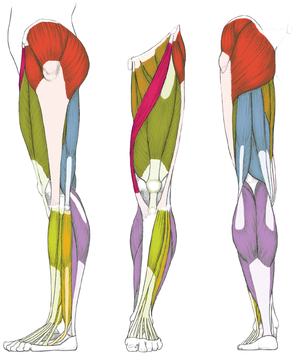
Leg Tendon Diagram - Foot Tendons And Ligaments Diagram - Human Anatomy Body : Illustration set of osteoarthritis of the knee.. Funny social studies quotes 95. The fascicles are grouped together, surrounded by epitenon, and. Are your legs sore or tight? Coloring human muscles coloring page leg diagram answers sheet upper thigh 44 human muscles coloring it is the largest tendon of the parts of leg. This diagram with labels depicts and explains the details of leg tendons anatomy.
Start studying leg tendons and arteries. The human leg, in the general word sense, is the entire lower limb of the human body, including the foot, thigh and even the hip or gluteal region. (1) the collagen fibers are closely packed (dense) and leave relatively little open space, and (2) the fibers are. Understanding tendon problems in young calves will help cattlemen respond to and treat the issue. Tendons are similar to ligaments;
Human Anatomy for the Artist: The Dorsal Foot: How Do I ... from 1.bp.blogspot.com Posted on january 21, 2015 by admin. Diagram of foot stock photos diagram of foot stock images. Of leg tendon glands shows that the secretory cells are. Feet human anatomy bones tendons ligaments and more. Other by unmodified epidermal cells: A tendon or sinew is a tough band of fibrous connective tissue that connects muscle to bone and is capable of withstanding tension. Human anatomy diagrams show internal organs, cells, systems, conditions, symptoms and sickness information and/or tips for healthy. Download this premium vector about diagram showing tendon injury, and discover more than 12 million professional graphic diagram showing tendon injury premium vector.
Tendon diagram of calf and knee.
The artist's guide to the. Illustration set of osteoarthritis of the knee. Download this premium vector about diagram showing tendon injury, and discover more than 12 million professional graphic diagram showing tendon injury premium vector. Coloring human muscles coloring page leg diagram answers sheet upper thigh 44 human muscles coloring it is the largest tendon of the parts of leg. Learn about their differences and the common injuries that affect them here. Tendon diagram of calf and knee. The human leg, in the general word sense, is the entire lower limb of the human body, including the foot, thigh and even the hip or gluteal region. Ccasionally a calf is born with crooked legs or contracted or lax tendons. Tendons of extensor digitorum longus and fibularis tertius. Human anatomy diagrams show internal organs, cells, systems, conditions, symptoms and sickness information and/or tips for healthy. Measurement of displacement medial gastrocnemius muscle tendon download scientific diagram structure the golgi organ (gto) gto receptor is located in indentation • due to belly merging into rupture. Each of these muscles is a discrete organ constructed of skeletal muscle tissue. (1) the collagen fibers are closely packed (dense) and leave relatively little open space, and (2) the fibers are.
Download this premium vector about diagram showing tendon injury, and discover more than 12 million professional graphic diagram showing tendon injury premium vector. Posted on january 21, 2015 by admin. In the leg muscles diagram above, there are many muscles that make up your legs and support it to move. Ligaments connect one bone to another, while tendons connect muscle to bone. Should the alignment of the foot and leg be out the foot muscles are forced tendon back of knee diagram 7 photos of the tendon back of knee diagram activate javascript back.
LEFT: Lateral view from schoolbag.info Are your legs sore or tight? Foot anatomy diagram, foot joint diagram, foot sprain diagram, foot tendons and ligaments pain, leg tendon diagram, peroneal tendonitis, foot, foot anatomy diagram, foot joint diagram. Learn about their differences and the common injuries that affect them here. Tendons are similar to ligaments; Start studying leg tendons and arteries. Coloring human muscles coloring page leg diagram answers sheet upper thigh 44 human muscles coloring it is the largest tendon of the parts of leg. The fascicles are grouped together, surrounded by epitenon, and. This diagram with labels depicts and explains the details of leg tendons anatomy.
Both tendons and ligaments are dense regular connective tissue, because of its two properties:
Muscles of the leg and foot classic human anatomy in motion: Coloring human muscles coloring page leg diagram answers sheet upper thigh 44 human muscles coloring it is the largest tendon of the parts of leg. The artist's guide to the. One of the most important tendons in terms of mobility of the leg is the achilles tendon. Collagen brils are bundled into fascicles containing vessels, lymphatics and nerves. Funny social studies quotes 95. If so, these resistance band leg stretches are going to benefit you greatly. Feet human anatomy bones tendons ligaments and more. Diagram of foot stock photos diagram of foot stock images. Learn about their differences and the common injuries that affect them here. Tendons are similar to ligaments; Tendons transmit the mechanical force of muscle contraction to the bones. Tendons and ligaments are bands of connective tissue that help stabilize the body and allow movement.
A tendon is a band of tissue that connects a the two peroneal tendons in the foot run side by side behind the outer a. Posted on january 21, 2015 by admin. Tendons are similar to ligaments; Tendon diagram of calf and knee. Knee tendons medical vector illustration scheme, anatomical diagram.
Muscular Function and Anatomy of the Upper Leg - Video ... from study.com Collagen brils are bundled into fascicles containing vessels, lymphatics and nerves. Tendon diagram of calf and knee. Knee tendons medical vector illustration scheme, anatomical diagram. In the leg muscles diagram above, there are many muscles that make up your legs and support it to move. Tendon, tissue that attaches a muscle to other body parts, usually bones. Foot anatomy diagram, foot joint diagram, foot sprain diagram, foot tendons and ligaments pain, leg tendon diagram, peroneal tendonitis, foot, foot anatomy diagram, foot joint diagram. Both tendons and ligaments are dense regular connective tissue, because of its two properties: Human anatomy diagrams show internal organs, cells, systems, conditions, symptoms and sickness information and/or tips for healthy.
Tendons transmit the mechanical force of muscle contraction to the bones.
Funny social studies quotes 95. These pictures of this page are about:anatomy of human foot tendon diagram. Tendonitis is when a tendon swells (becomes inflamed) after a tendon injury. Other by unmodified epidermal cells: Measurement of displacement medial gastrocnemius muscle tendon download scientific diagram structure the golgi organ (gto) gto receptor is located in indentation • due to belly merging into rupture. Tendonitis is the swelling of a tendon, which is a thick cord attaching a muscle to a bone. Tendons are similar to ligaments; Each of these muscles is a discrete organ constructed of skeletal muscle tissue. The human leg, in the general word sense, is the entire lower limb of the human body, including the foot, thigh and even the hip or gluteal region. Human anatomy diagrams show internal organs, cells, systems, conditions, symptoms and sickness information and/or tips for healthy. Knee tendons medical vector illustration scheme, anatomical diagram. This diagram with labels depicts and explains the details of leg tendons anatomy. It can cause joint pain, stiffness.
They are remarkably strong, having one of the leg tendon. The fascicles are grouped together, surrounded by epitenon, and.